-

生物通官微
陪你抓住生命科技
跳动的脉搏
高内涵筛选在生命科学研究中的应用[创新技巧]
【字体: 大 中 小 】 时间:2011年07月08日 来源:赛默飞世尔科技
编辑推荐:
过去的几十年来,成像作为一种基于细胞的检测模式,在现有的和正在开发的生物模型中,开启了一个测量“终点表型”的全新世界。这些“高内涵”方法结合了多种细胞生理学的测量手段,不管它是来源于亚细胞组分、多细胞结构还是模式生物。这样产生的多层面的数据可以帮助人们对很多复杂现象产生新的认知,这些现象可以包括细胞分化,也可以包括化合物药物学和毒理学研究。我们查阅了日益增加的文献报道,了解其主要的应用领域,已发现了充足的证据表明这项科学技术正在对药物研发和生命科学产生着实实在在的影响。
Joe M. Zock
赛默飞世尔科技公司 美国
关键字:高内涵筛选;细胞信号通路;肿瘤学;神经生物学;体外毒理学;靶点鉴定;靶点验证;RNA干扰;干细胞
简介
高内涵筛选(High Content Screening, HCS)是自动化显微成像技术与图像分析技术在药物研发及细胞生物学方面的一个应用。这个技术已经从最初的一个有趣命题发展成为一种有用的技术,并在最近十年中演变成为有价值的实用方法。这篇综述旨在总结那些发表在同行审阅的期刊中,并使用HCS作为一个主要研究方法的文章,试图让大家了解HCS在生命科学的几个重要领域中的广泛应用。
可以想见,早期的研究论文着重于把HCS作为一种新技术来发表。当这一技术得到广泛的接受和使用时,研究的侧重点又回到了被研究的生物学问题,而HCS则成为一种工具,为任何新化合物或新假设提供“支持性的生物学证据”。作者认为,这种趋势是一项新技术被真正采纳的标志,也是HCS这项技术被保留下来的有力证明。
有趣的是,虽然大多数使用HCS的论文可归类为细胞信号通路、肿瘤学、神经生物学、体外毒理学及靶点验证(例如,RNAi),然而越来越多的新近发表的文章则描述了HCS全新的应用。在上述“标准”应用之外的领域,HCS技术已被广泛接受,体现了这种技术的灵活性,可以扩展到其它可研究的生物学过程,包括心脏衰竭[1],间隙连接[2],免疫抑制[3],骨质疏松[4],吞噬作用[5],自我吞噬作用[6],中心粒功能[7],真菌致病机制[8],视网膜修复[9],生理节律[10],以及酵母筛查[11]等等,不一而足。
像其它新技术一样,HCS也被拿来与当前的技术实验方法做比较。大多数情况下,HCS相比其它技术手段具有显著的优势;或至少被认为是与现有技术互补的,可以提供更多的数据来用于得出科学结论。例如,在最近将HCS与标准酶联免疫吸附实验(ELISA)相比较的文章中,可以看到HCS可以从单个细胞的水平来观察细胞内正在发生的事件,使得测量结果更准确和可靠[12,13]。
索取Cellomics ArrayScan HCS平台的更多资料
Liu等解释了ELISA和HCS如何提供互补的结果,来帮助作者确定受试化合物的复杂的药理学原理。文中提到:“很清楚,神经微丝蛋白(NF)的ELISA结果是对神经细胞活性的评估,…… 另一方面,Cellomics ArrayScan HCS平台,象本研究中所展示的,旨在评估化合物促进神经突触生长的能力,……两种技术平台结合使用,能够帮助研究人员鉴定出具有多种细胞活性的化合物,例如FK506”[14]。
Aglei等引证了使用HCS检测蛋白/蛋白相互作用的亚细胞水平定位,这是HCS优于第二信使检测的一个方面。HCS能够识别一个细胞培养孔内的某一特定细胞群,使得瞬时转染能够被用于可靠的筛选研究,不再需要强制性地使用稳定转染技术,因为基因的过度表达可以导致异常的生理学和毒理学变化。“HCS实验提供了其它信息,这些信息不可能从荧光偏振(FP)和报告基因实验中获得,例如蛋白/蛋白相互作用发生的亚细胞定位。”在这种实验筛选策略下,与购买FP和报告基因试剂的价格相比,HCS转位检测可以称得上是一种价格低廉的实验方案[15]。
越来越多的困难实验正在被HCS所取代,这样做的好处可能有:比现有方法的灵敏度更高,提高了通量,增加了安全性,和/或降低了成本。Baniecki等人将HCS用于寻找抗疟新药,他认为,“使用基于图像的DAPI恶性疟原虫生长实验,我们可以检测到单个疟原虫;而使用DAPI恶性疟原虫生长实验和[3H]标记的次黄嘌呤实验,96孔板上得到的读数结果显示为:具有活力的疟原虫的比例为0.25%。两种手段的结果相比较,HCS的灵敏度及可靠性都显著提高[16]。疾病控制中心的Johnson等建立了一个快速的高通量牛痘病毒中和实验[17],此方法应用HCS来取代那些“费力的检测方法,尤其是当待检样品数量很多时,需要48-72小时来等待空斑的形成,还需要更多的时间来分析结果,结果很可能带有很大的主观性-因为菌斑是手工计数的。”HCS检测的是带有GFP的病毒感染,“因其生成数据的速度和可靠性,具有取代空班减少中和滴度的可能,而成为临床实验室进行中和检测的标准方法。在正痘病毒爆发事件中,HCS的快速和高通量的特性已证明了它是非常有价值的。
从历史角度看,HCS源自药物发现,使用此方法可检测多个参数,同时又可检测单个细胞的特性,因此,该方法起初被用做新型的复筛检测模式、选择性筛选和细胞毒性的特征描绘。“高内涵”、“前后关联”和“具有相关性”等词语描述了出自于当前的HCS平台的数据特点[18],但是,理解这些数据的价值和应用范围仅在近期的文献报道中才变得越来越多。一个非常显著的例子是,Young等人在他们最近的文章中,将HCS数据和配体-靶点预测相结合,探讨化合物的药理学机制,非常精彩地表明了应用多层面的HCS数据是如何的强大[19]。使用细胞周期排列作为模型,收集到了一系列的基于图像的细胞学参数。实验选择的是带有36特征的亚集,选定了六个参数(胞核大小、复制、有丝分裂、胞核形态、EdU纹理和胞核椭圆度),用于对拥有6547个化合物的库进行了特征性描绘。“根据反应参数,有活性的化合物被归为7个表型效应的类别中。然后,我们探索了表型特征与活性化合物的结构之间的关系,并预测了目标化合物的结构特征。这样所产生的构效关系比仅用单一类型的数据所产生的结果要丰富得多,而且,我们可以推断一些化合物的作用机制。”
HCS与细胞信号通路
在药物研发的过程中,理解环境诱发因素如何触发一个特定的生物级联反应是找到有效治疗方法的关键。因此,细胞信号通路是开发大多数特异性作用于靶点的药物的基础。在科研领域,同样存在着这样的需求―研究新发现的蛋白是如何参与到各种各样的信号通路的。因此,在同行评审的期刊中,常常可以看到引用HCS的文章报道的是细胞信号转导的某些领域,就不足为奇了。从十年前最初的HCS文章报道NFkB转位[20],许多信号分子的活性已被量化,包括STAT[21,22],wnd/fzd[23],akt[24],NFAT[25],p38[26],TGF-beta[27]和Smad2/3[28,29],这组成了包括从炎症反应[30]到G-蛋白偶联受体[31-33]的信号通路网络。
HCS与肿瘤学
通过早期检测凋亡[34-38]和增殖[39-41],HCS在肿瘤研究领域找到了最初的立足点。接着, 关于细胞周期[42-44],转化[45]和迁移[46-48]的实验随之而来。我们开发了迁移实验算法和试剂盒,用来评价肿瘤转移的可能性[49-51]。利用HCS能够看到单个细胞的应答,而不是“群体的平均反应性”,这使我们能够更好地理解抗肿瘤化合物是怎样对癌变细胞和正常细胞产生不同的作用的,而且,也能够帮助我们确定肿瘤标志物的功能[52]。使用HCS所鉴定的抗肿瘤化合物已经开始进入临床实验阶段[53]。
随着HCS技术的不断发展,它也逐渐出现在肿瘤研究的其它领域。例如血管生成,现在已经成为一项常规操作,在微孔板中,通过刺激内皮细胞,使其经历向生成血管的状态转化[54,55]。许多细胞相互传递信号,一起协同工作,产生具有特别功能的多细胞结构,这些多细胞结构各自具有不同的特性,分别与不同的疾病状态相关联,在这一点上,血管生成做为一个检测指标是非常令人兴奋的。刺激新血管生成,对于受损器官或伤口而言,可能是有利的;而抑制新血管生成,对于实体瘤或视网膜(湿型黄斑部变性)而言,则可能具有治疗作用。这里的关键是要能够准确地捕捉、测量和显示这些表型,这是需要通过对很多参数逐个检测才能得到的。在血管生成这个例子中,能够测量血管的大小和形状、连结度、节点的个数、血管中细胞的个数、甚至血管中细胞的靶点激活情况,可帮助研究者辨别不同的化合物的活性。在肿瘤研究中使用HCS的另一个热点领域是:通过定量检测细胞骨架的重排,特别是微管的组装和解体,来评价化合物的抗肿瘤作用[56,57]。
NCI化学基因组研究所是充分应用HCS平台的一个非常好的例子。他们建立和应用HCS检测方法进行了核点形成、细胞形态改变以及蛋白转位的研究。“因为这种检测结果是在细胞水平完成的,而不是一个板孔内的平均状况,信噪比非常高;本质上,每一个板孔本身就是一套数据点”[58]。利用HCS,他们鉴定出了新型细胞分裂调节剂以及其它几种涉及NFAT和FOX01a核转位的调节因子,这是通过检测多种细胞运动模式来完成的,而不是像研究经典抗有丝分裂化合物那样,仅粗略检测微管的破裂。这里提到的经典的抗有丝分裂化合物,指的是secramine,它是一种肌动蛋白多聚体的抑制剂,可减少肿瘤转移。
HCS与神经生物学
在HCS技术发展的最早期,我们就认识到了成像技术在神经细胞形态的量化方面具有非常大的潜力,我们是最早开发出产品来监测神经轴突生长的机构。多年来,根据许多进行神经轴突生长刺激剂[59-62]和神经保护[63-65]研究的用户的反馈,我们对算法进行了很多次的优化,使得结果中计算出的指标在原代细胞和标准细胞系中都能够代表神经元和神经亚群体的许多特性。
更令人激动的是,不管是对潜在的基本生物学原理的研究、还是建立新模型、或是为了治疗干预而进行的药物分子筛选,HCS在多种神经疾病状态的研究中已被广泛应用。这样的例子包括Alzheimer氏病[66],Parkinson氏病[67,68],Huntington氏病[69,70],肌萎缩性侧索硬化[71]和脑肿瘤[72],每年都有更多的文章出现。
HCS与体外毒理学
某种程度上,所有的HCS检测方法都可以认为是“毒性”分析,因为这些方法测量的是细胞对刺激的生理学应答,不管这种刺激来自环境还是化学物质。从相对简单的即刻细胞毒性检测,例如细胞计数及细胞圆度,到更为特异的细胞器健康状态的检测[73,74],HCS可以应用于多种情形,通常是作为多参数检测,这种情况下,将多个指标进行交叉关联分析,会帮助深入确定毒性状态。
很清楚,HCS已经在药物研发中细胞毒性分析领域确立了坚实的地位[75]。然而,超越于直接检测细胞毒作用的应用领域,即能够做到在细胞水平进行预测,来评估对于整个生物体(例如我们人类)产生的下游毒性效应,这才是药物发现中自动化成像领域的重要发展方向,因为能否在正确的时间获取关键的数据,这种能力的不断增强就意味着上亿美元的得失。最初的应用领域包括:采用微核诱导检测[77,78]来评估基因毒性,检测肝脂肪沉积来评估是否患有磷脂质病[79],以及发育神经毒性[80]。展望未来,HCS技术在建立新型的毒性分析模型[81],包括使用模式生物如斑马鱼和线虫方面,还具有很大的潜力。
一个最令人兴奋的结果证明了多参数成像方法对于体外毒性学研究在药物诱导的肝损伤领域中的潜力。这里,Xu等对一系列的与肝毒性直接相关的表型建立了一个检测方案[82]。“在检测300多种药物和化合物(其中包括许多可以导致人类中罕见的肝毒性药物)时,使用我们的检测方案,检出率达到50-60%,假阳性率非常低,仅为0-5%”。
在另一个肝毒性化合物的追溯调查中,O’Brien等[83]将业内广泛使用的“标准7项”生化细胞毒性分析方法与一种4通道多参数的HCS分析方法进行了比较。结果表明,与“标准7项”生化分析中最佳组合项目相比较,HCS具有更高的灵敏度(93%比<25%)和特异性(98%比~90%)。这些研究成果在制药产业界得到了广泛证实[84]。
HCS与靶点验证
药物发现早期的靶点验证,以及大多数基础研究,都着眼于寻找细胞生物学架构中的新成员,以及验证它们各种各样的功能。在基础研究方面,靶点验证可以帮助理解细胞生物学的全景;在药物发现方面,靶点验证为开发反应疾病状态的检测分析方法提供了基础,从而可以找到阻止疾病状态的分子。最终,这个领域的成功与否取决于相关性高的生物学模型及准确的生理环境条件。相对较晚出现的技术是利用干细胞和RNAi来产生细胞模型,这是一个对表型进行量化检测的绝佳手段。不管是追踪一个正在分化的干细胞变成肌细胞群的发展过程,还是评估敲除神经轴突细胞生长的增殖信号的结果,HCS都可应用于其中。在最近的发表一篇基于RNAi筛选的综述中,Perrimon评论到,“在RNAi HTS中也许最显著的进展将会来自高内涵筛选。依赖细胞表型的HCS检测在RNAi HTS中正在成为一种首选的方法,因为它们产生的数据集信息丰富,原代细胞的使用提供了很多机会,能够在生物相关性比较高的环境下,对细胞形态进行筛选”[85]。
Moffat等建立了一种基于高内涵成像的筛选方法,来鉴定有丝分裂进程中所必须的基因,并针对5000种表达独特shRNA、以1028个人类基因为靶点的慢病毒载体进行了筛选[86]。这个筛选找到了大约100个(新型)增殖相关的候选调控元件。类似的利用HCS筛选siRNA文库来监测表型参数的研究项目也已经完成[87-90]。
在干细胞研究的前沿,HCS已被用于帮助鉴定与干细胞自我更新[91,92]和分化[93]相关的调节系统,主要是通过胚胎干细胞[94,95]或从成年人组织衍生出的细胞系[96]中的多能性标记物(例如,Oct-4)进行量化检测。通过使用多种分化状态的生物标记物,已经完成了对细胞命运的下游追踪[97]。 Peerani等阐明了HCS具有可以提供细胞与细胞的空间关系的独特能力,该研究揭示了人胚胎干细胞(hESC)培养体系的异质微环境(小生态环境)对hESC的命运产生了影响[98]。将细胞分化的诱导和抑制因子(通过siRNA下调)的分泌局限化以后,通过检测niche的大小和细胞组成,“我们率先发现了Smad1在空间信息整合、以及niche大小相关的hESC自我更新和分化中的控制作用。”
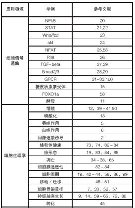
表1.HCS主要的应用领域及参考文献举例

未来的方向及展望
从上述引用的文献中,可以清楚地看到HCS已经超越了“证明自身技术”的阶段,而正在进入“被广泛采用”的阶段,随着用户需求的不断增加,将推动这项技术变得更加节约成本、更易于使用,并且更加可靠。新试剂[99]和微环境的开发,与自发荧光蛋白的持续应用[100]一起,将打开一扇通往更多量化检测细胞行为的大门。着眼于HCS的生产力指标,产生信息(分析前处理自动化,试剂盒,算法)和理解这些信息(数据管理,数据可视化,数据挖掘)两方面都将成为对检测平台的重要补充,使得HCS产生的常规数据成为生命科学研究中支持决策的一部分证据。随着HCS从原来的主要应用范围向大规模使用演化,技术开发将通过使用多细胞集群、组织、器官和有机体向加强生物数据相关性迈进。另一个趋势是:将细胞组学与基因组学、蛋白组学技术结合起来,将会提供一个前所未有的细胞功能的蓝图。
由于许多国家正在提议减少动物实验,因此,在不远的将来,HCS的前景是非常光明的[101,102]。现在,正是开发细胞分析工具(诸如HCS)的最佳时机。作为一个从一开始就参与开发此技术的人员,我可以诚实地说,由于一些“高产出用户”的热情和贡献,HCS作为一个强大的工具,正在贡献它的力量,帮助构建未来的“科学知识架构”。
索取Cellomics ArrayScan HCS平台的更多资料
参考文献
[1] Harrison, B.C.; Kim, M.S., van Rooji, E.; Plato, C.F.; Papst, P.J.; Vega, R.B.; McAnally, J.A.; Richardson, J.A.; Bassel-Duby, R.; Olson, E.N.; McKinsey, T.A. Regulation of cardiac stress signaling by protein kinase D1. Mol Cell. Biol., 2006, 26(10), 3875-3888.
[2] Li, Z.; Yan, Y.; Powers, E.A.; Ying, X.; Janjua, K.; Garyantes, T.; Baron, B. Identification of gap junction blockers using automated fluorescence microscopy imaging. J. Biomol. Screen. 2003, 8, 489-499.
[3] Yan, P.; Nanamori, M.; Sun, M.; Zhou, C.; Cheng, N.; Li, N.; Zheng, W.; Xiao, L.; Xie, X.; Ye, R.D.; Wang, M.W. The immunosuppressant cyclosporin A antagonizes human formyl peptide receptor through inhibition of cognate ligand binding. J. Immunol., 2006, 177, 7050-7058.
[4] Buckbinder, L.; Crawford, D.T.; Qi, H.; Ke, H.Z.; Olson, L.M.; Long, K.R.; Bonnette, P.C.; Baumann, A.P.; Hambor, J.E.; Grasser, W.A.; Pan, L.C.; Owen, T.A.; Luzzio, M.J.; Hulford, C.A.; Gebhard, D.F.; Paralkar, V.M.; Simmons, H.A.; Kath, J.C.; Roberts, W.G.; Smock, S.L.; Guzman-Perez, A.; Brown, T.A.; Li, M. Proline-rich tyrosine kinase 2 regulates osteoprogenitor cells and bone formation and offers an anabolic treatment approach for osteoporosis. Proc. Natl. Acad. Sci., USA, 2007, 104(25), 10619-10624.
[5] Steinberg, B.E.; Scott, C.C.; Grinstein, S. High-throughput assays of phagocytosis, phagosome maturation, and bacterial invasion. Am. J. Physiol. Cell. Physiol., 2007, 292, 945-952.
[6] Zhang, L.; Yu, J.; Pan, H.; Hu, P.; Hao, Y.; Cai, W.; Zhu, H.; Yu, A.D.; Xie, X.; Ma, D.; Yuan, J. Small molecule regulators of autophagy identified by an image-based high-throughput screen. Proc. Natl. Acad. Sci. USA, 2007, 104(48), 19023-19028.
[7] Rogers, G.C.; Rusan, N.M.; Peifer, M.; Rogers, S.L. A Multicomponent assembly pathway contributes to the formation of acentrosomal microtubule arrays in interphase drosophila cells. Mol. Biol. Cell., 2008, 19, 3163-3178.
[8] Wheeler, R.T.; Fink, G.R. A drug-sensitive genetic network masks fungi from the immune system. PLoS Pathog., 2006, 2(4), e35 328-339.
[9] Kerrison, J.B.; Lewis, R.N.; Otteson, D.C.; Zack, D.J. Bone morphogenic proteins promote neurite outgrowth in retinal ganglion cells. Mol. Vision, 2005, 11, 208-215.
[10] Badura, L.; Swanson, T.; Adamowicz, W.; Adams, J.; Cianfrogna, J.; Fisher, K.; Holland, J.; Kleiman, R.; Nelson, F.; Reynolds, L.; St. Germain, K.; Schaeffer, E.; Tate, B.; Sprouse, J. An inhibitor of casein kinase I induces phase delays in circadian rhythms under free-running and entrained conditions. J. Pharmacol. Exp. Ther.,
2007, 322, 730-738.
[11] Benanti, J.A.; Cheung, S.K.; Brady, M.C.; Toczyski, D.P. A proteomic screen reveals SCF[Grr1] targets that regulate the glycolytic-gluconeogenic switch. Nat. Cell Biol., 2007, 9(10), 1184-1191.
[12] Gasparri, F.; Mariani, M.; Sola, F.; Galvani, A. Quantification of the proliferation index of human dermal fibroblast cultures with the ArrayScan high content screening reader. J. Biomol. Screen. 2004, 9, 232-243.
[13] Vogt, A.; Cooley, K.A.; Brisson, M.; Tarpley, M.G.; Wipf, P.; Lazo, J.S. Cell-active dual specificity phosphatase inhibitors identified by high content screening. Chem. Biol., 2003, 10, 733-742.
[14] Liu, D.; McIlvain, H.B.; Fennell, M.; Dunlop, J.; Wood, A.; Zaleska, M.M.; Graziani, E.I.; Pong, K. Screening of immunophilin ligands by quantitative analysis of neurofilament expression and neurite outgrowth in cultured neurons and cells. J. Neurosci. Methods, 2007, 163, 310-320.
[15] Agler, M.; Prack, M.; Zhu, Y.; Kolb, J.; Nowak, K.; Ryseck, R.; Shen, D.; Cvijic, M.E.; Somerville, J.; Nadler, S.; Chen, T. A high content glucocorticoid receptor translocation assay for compound mechanism-of-action evaluation. J. Biomol. Screen., 2007, 12, 1029-1041.
[16] Baniecki, M.L.; Wirth, D.F.; Clardy, J. High-Throughput Plasmodium falciparum growth assay for malaria drug discovery. Antimicrob. Agents Chemother., 2007, 51(2), 716-723.
[17] Johnson, M.C.; Damon, I.K.; Karem, K.L. A Rapid, highthroughput vaccinia virus neutralization assay for testing smallpox vaccine efficacy based on detection of green fluorescent protein. J. Virol. Methods, 2008, 150, 14-20.
[18] Keefer, S.; Zock, J. In Approaching High Content Screening and Analysis: Practical Advice for Users; Haney, S.A., Eds.; High Content Screening: Science, Techniques and Applications. Wiley-Interscience, 2008, pp. 3-24.
[19] Young, D.W.; Bender, A.; Hoyt, J.; McWhinnie, E.; Chirn, G.W.; Tao, C.Y.; Tallarico, J.A.; Labow, M.; Jenkins, J.L.; Mitchison, T.J.; Feng, Y. Integrating high content screening and ligand-target prediction to identify mechanism of action. Nat. Chem. Biol., 2008, 4, 59-68.
[20] Ding, G.J.F.; Fischer, P.A.; Boltz, R.C.; Schmidt, J.A.; Colaianne, J.J.; Gough, A.; Rubin, R.A.; Miller, D.K. Characterization and quantitation of NF-kB nuclear translocation induced by interleukin-1 and tumor necrosis factor-a. J. Biol. Chem., 1998, 273, 28897-28905.
[21] Vakkila, J.; DeMarco, R.A.; Lotze, M.T. Imaging analysis of STAT1 and NF-kB translocation in dendritic cells at the single cell level. J. Immunol. Methods, 2004, 294, 123-134.
[22] Grace, M.J.; Lee, S.; Bradshaw, S.; Chapman, J.; Spond, J.; Cox, S.; DeLorenzo, M.; Brassardi, D.; Wylie, D.; Cannon-Carlson, S.; Cullen, C.; Indelicato, S.; Voloch, M.; Bordens, R. Site of pegylation and polyethylene glycol molecule size attenuate inferferon-alpha antiviral and antiproliferative activities through the JAK/STAT signaling pathway. J. Biol. Chem., 2004, 280(8), 6327-6336.
[23] Borchert, K.M.; Galvin, R.J.; Frolik, C.A.; Hale, L.V.; Halladay, D.L.; Gonyier, R,J.; Trask, O.J.; Nickischer, D.R.; Houck, K.A. High content screening assay for activators of the wnt/fzd pathway in primary human cells. Assay Drug Dev. Technol., 2005, 3(2), 133-141.
[24] Lundholt, B.K.; Linde, V.; Loechel, F.; Pedersen, H.C.; Moller, S.; Praestegaard, M.; Mikkelsen, I.; Scudder, K.; Bjorn, S.P.; Heide, M.; Arkhammar, P.O.; Terry, R.; Nielsen, S.J. Identification of Akt pathway inhibitors using Redistribution® screening on the FLIPR and the InCell 3000 Analyzer. J. Biomol. Screen., 2005, 10, 20-29.
[25] Van Sant, C.; Wang, G.; Anderson, M.G.; Trask, O.J.; Lesniewski, R.; Semizarov, A. Endothelin signaling in osteoblasts: global genome view and implication of the calcineurin/NFAT pathway. Mol. Cancer Ther., 2007, 6(1), 253-261.
[26] Ross, S.; Chen, T.; Yu, V.; Tudor, Y.; Zhang, D.; Liu, L.; Tamayo, N.; Dominguez, C.; Powers, D. High content screening analysis of the p38 pathway: profiling of structurally related p38alpha kinase inhibitors using cell-based assays. Assay Drug. Dev. Technol., 2006, 4(4), 397-409.
[27] Sarker, K.P.; Kataoka, H.; Chan, A.; Netherton, S.J.; Pot, I.; Huynh, M.A.; Feng, X.; Bonni, A.; Riabowol, K.; Bonni, S. ING2 as a novel mediator of transforming growth factor-β-dependent responses in epithelial cells. J. Biol. Chem., 2008, 283, 13269-13279.
[28] Dawes, L.J.; Angell, H.; Sleeman, M.; Reddan, J.R.; Wormstone, I.M. TGFb isoform dependant Smad2/3 kinetics in human lens epithelial cells: a cellomics analysis. Exp. Eye Res., 2007, 84, 1009-1012.
[29] Wormstone, I.M.; Dawes, L.J.; Sleeman, M.; Anderson, I.K.; Reddan, J.R. FGF promotes TGF-beta-induced matrix contraction and suppresses expression of the transdifferentiation marker alphaSMA. Invest. Ophthalmol. Vis. Sci., 2008, 49, 3726.
[30] Bertelsen, M.; Sanfridson, A. Inflammatory pathway analysis using a high content screening platform. Assay Drug Dev. Technol.,2005, 3, 261-271.
[31] Schlag, B.D.; Lou, Z.; Fennell, M.; Dunlop, J. Ligand dependency of 5-HT2C receptor internalization. J. Pharmacol. Exp. Ther., 2004, 310, 865-870.
[32] Fowler, A.; Seifert, N.; Acker, V.; Woehrle, T.; Kilpert, C.; de Saizieu, A. A nonradioactive high-throughput / high content assay for measurement of the human serotonin reuptake transporter function in vitro. J. Biomol. Screen. 2006, 11, 1027-1034.
[33] Barnard, R.; Barnard, A.; Salmon, G.; Liu, W.; Sreckovic, S. Histamine-induced actin polymerization in human eosinophils: an imaging approach for histamine H4 receptor. Cytometry A, 2008, 73(4), 299-304.
[34] Inglefield, J.R.; Larson, C.J.; Gibson, S.J.; Lebrec, H.; Miller, R.L. Apoptotic responses in squamous carcinoma and epithelial cells to small-molecule toll-like receptor agonists evaluated with automated cytometry. J. Biomol. Screen., 2006, 11(6), 575-585.
[35] Lovbor, H.; Gullbo, J.; Larsson, R. Screening for apoptosisclassical and emerging techniques. Anticancer Drugs, 2005, 16, 593-599.
[36] To, K.; Zhao, Y.; Jiang, H.; Hu, K.; Wang, M.; Wu, J.; Lee, C.; Yokom, W.; Stratford, A.L.; Klinge, U.; Mertens, P.R.; Chen, C.S.; Bally, M.; Yapp, D.; Dunn, S.E. The phosphoinositide-dependent kinase-1 inhibitor 2-amino-N-[4-[5-(2-phenanthrenyl0-3-(trifluoromethyl)-1hH-pyrazol-1-yl]phenyl]-acetamide (OSU-03012) prevents Y-box binding protein-1 from inducing epidermal growth factor receptor. Mol. Pharmacol., 2007, 72(3), 641-652.
[37] Vogt, A.; McDonald, P.R.; Tamewitz, A.; Sikorski, R.P.; Wipf, P.; Skoko, III J.J.; Lazo, J.S. A cell-active inhibitor of mitogenactivated protein kinase phosphatases restores paclitaxel-induced apoptosis in dexamethasone-protected cancer cells. Mol. Cancer Ther., 2008, 7, 330-340.
[38] Cummings, J.; Hodgkinson, C.; Odedra, R.; Sini, P.; Heaton, S.P.; Mundt, K.E.; Ward, T.H.; Wilkinson, R.W.; Growcott, J.; Hughes, A.; Dive, C. Preclinical evaluation of M30 and M65 ELISAs as biomarkers of drug induced tumor cell death and antitumor activity. Mol. Cancer Ther., 2008, 7, 455-463.
[39] Soncini, C.; Carpinelli, P.; Gianellini, L.; Fancelli, D.; Vianello, P.; Rusconi, L.; Storici, P.; Zugnoni, P.; Pesenti, E.; Croci, V.; Ceruti, R.; Giorgini, M.L.; Cappella, P.; Ballinari, D.; Sola, F.; Varasi, M.;
Bravo, R.; Moll, J. PHA-680632, a novel Aurora kinase inhibitor with potent antitumoral activity. Clin. Cancer Res., 2006, 12(13), 4080-4089.
[40] Gasparri, F.; Mariani, M.; Sola, F.; Galvani, A. Quantification of the proliferation index of human dermal fibroblast cultures with the ArrayScan high content screening reader. J. Biomol. Screen., 2004, 9, 232-243.
[41] Breier, J.M.; Radio, N.M.; Mundy, W.R.; Shafer, T.J. Development of a high-throughput screening assay for chemical effects on proliferation and viability of immortalized human neural progenitor cells. Toxicol. Sci., 2008, 105, 119-133.
[42] Barabasz, A.; Foley, B.; Otto, J.C.; Scott, A.; Rice, J. The use of high content screening for the discovery and characterization of compounds that modulate mitotic index and cell cycle progression by differing mechanisms of action. Assay Drug Dev. Technol., 2006, 4(2), 153-163.
[43] Gasparri, F.; Ciavolella, A.; Galvani, A. Cell cycle inhibitor profiling by high content analysis. Adv. Exp. Med. Biol., 2007, 604, 137-148.
[44] Gasparri, F.; Cappella, P.; Galvani, A. Multiparametric cell cycle analysis by automated microscopy. J. Biomol. Screen., 2006, 11, 586-598.
[45] Li, Y.; Pan, J.; Li, J.L.; Lee, J.H.; Tunkey, C.; Saraf, K.; Garbe, J.C.; Whitley, M.Z.; Jelinsky, S.A.; Stampfer, M.R.; Haney, S.A. Transcriptional changes associate with breast cancer occur as normal human mammary epithelial cells overcome senescence barrier and become immortalized. Mol. Cancer, 2007, 6(7), 1-17.
[46] Mastyugin, V.; McWhinnie, E.; Labow, M.; Buxton, F. A quantitative high-throughput endothelial cell migration assay. J. Biomol. Screen., 2004, 9, 712-718.
[47] Richards, G.R.; Millard, R.M.; Leveridge, M.; Kerby, J.; Simpson, P.B. Quantitative assays of chemotaxis and chemokinesis for human neural cells. Assay Drug Dev. Technol., 2004, 2, 465-472.
[48] Nam, J.-S.; Ino, Y.; Kanai, Y.; Sakamoto, M.; Hirohashi, S. 5-aza-2'-deoxycytidine restores the E-cadherin system in E-cadherinsilenced cancer cells and reduces cancer metastasis. Clin. Exp. Metastasis, 2004, 21, 49-56.
[49] Bhawe, K.M.; Blake, R.A..; Clary, D.O.; Flanagan, P.M. An automated image capture and quantitation approach to identify proteins affecting tumor cell proliferation. J. Biomol. Screen. 2004, 9, 216-222.
[50] Shimamra, T.; Yasuda, J.; Ino, Y.; Gotoh, M.; Tsuchiya, A.; Nakajima, A.; Sakamoto, M.; Kanai, Y.; Hirohashi, S. Dysadherin expression facilitates cell motility and metastatic potential of human pancreatic cancer cells. Cancer Res., 2004, 64, 6989-6995.
[51] Chuma, M.; Sakamoto, M.; Yasuda, J.; Fujii, G.; Nakanishi, K.; Tsuchiya, A.; Ohta, T.; Asaka, M.; Hirohashi, S. Overexpression of cortactin is involved in motility and metastasis of hepatocellular carcinoma. J. Hepatol., 2004, 41, 629-636.
[52] O'Brien, C.; Cavet, G.; Pandita, A.; Hu, X.; Haydu, L.; Mohan, S.; Toy, K.; Rivers, C.S.; Modrusan, Z.; Amler, L.C.; Lackner, M.R. Functional genomics identifies ABCC3 as a mediator of taxane resistance in HER2-amplified breast cancer. Cancer Res., 2008, 68, 5380-5389.
[53] Sartore-Bianchi, A.; Gasparri, F.; Galvani, A.; Nici, L.; Darnowski, J.W.; Barbone, D.; Fennell, D.A.; Gaudino, G.; Porta, C.; Mutti, L. Bortezomib inhibits nuclear factor-kappa B dependent survival and
has potent in vivo activity in mesothelioma. Clin. Cancer Res., 2007, 13(19), 5942-5951.
[54] Liu, L.; Cao, Y.; Chen, C.; Zhang, X.; McNabola, A.; Wilkie, D.; Wilhelm, S.; Lynch, M.; Carter, C. Sorafenib blocks the RAF/MEK/ERK pathway, inhibits tumor angiogenesis, and induces tumor cell apoptosis in hepatocellular carcinoma model PLC/PRF/5. Cancer Res., 2006, 66(24), 11851-11858.
[55] Basu, P.; Ghosh, R.; Grove, L.; Klei, L.; Barchowsky, A. Angiogenic potential of 3-nitro-4-hydroxy benzene arsonic acid (Roxarsone). Environ. Health Perspect., 2008, 114(4), 520-523.
[56] Wei, S.Y.; Li, M.; Tang, S.A.; Sun, W.; Xu, B.; Cui, J.R.; Lin, W.H. Induction of apoptosis accompanying with G1 phase arrest and microtubule disasembly in human hepatoma cells by jaspolide B, a new isomalabaricane-type triterpene. Cancer Lett., 2008, 262, 114-122.
[57] Minguez, J.M.; Giuliano, K.A.; Balachandran, R.; Madiraju, C.; Curran, D.P.; Day, B.W. Synthesis and high content cell-based profiling of simplified analogues of the microtubule stabilizer (+)discodermolide. Mol. Cancer Ther., 2002, 1, 1305-1313.
[58] Tolliday, N.; Clemens, P.A.; Ferraiolo, P.; Koehler, A.N.; Lewis, T.A.; Li, X.; Schreiber, S.L.; Gerhard, D.S.; Eliasof, S. Small molecules, big players: the National Cancer Institute’s initiative for chemical genetics. Cancer Res., 2006, 66(18), 8935-8942.
[59] Richards, G.R.; Smith, A.J.; Parry, F.; Platts, A.; Chan, G.K.; Leveridge, M.; Kerby, J.E.; Simpson, P.B. A morphology-and kinetics-based cascade for human neural cell high content screening. Assay Drug Dev. Technol., 2006, 4(2), 143-152.
[60] McIlvain, H.B.; Baudy, A.; Sullivan, K.; Liu, D.; Pong, K.; Fennell, M.; Dunlop, J. Pituitary adenylate cyclase-activating peptide (PACAP) induces differentiation in the neuronal F11 cell line through a PKA-dependent pathway. Brain Res., 2006, 1077(1), 16-23.
[61] Loh, S.H.; Francescut, L.; Lingor, P.; Bahr, M.; Nicotera, P. Identification of new kinase clusters required for neurite outgrowth and retraction by loss-of-function RNA interference screen. Cell Death Differ., 2008, 15(2), 283-298.
[62] Qian, M.D.; Zhang, J.; Tan, X.Y.; Wood, A.; Gill, D.; Cho, S. Novel agonist monoclonal antibodies activate TrkB receptors and demonstrate potent neurotrophic activities. J. Neurosci., 2006, 26, 9394-9403.
[63] Zhou, W.; Zhu, X.; Zhu, L.; Cui, Y.Y.; Wang, H.; Qi, H.; Ren, Q.S.; Chen, H.Z. Neuroprotection of muscarinic receptor agonist pilocarpine against glutamate-induced apoptosis in retinal neurons. Cell. Mol. Neurobiol., 2008, 28, 263-275.
[64] Ruan, B.; Pong, K.; Jow, F.; Bowlby, M.; Crozier, R.A.; Liu, D.; Liang, S.; Chen, Y.; Mercado, M.L.; Feng, X.; Bennett. F.; von Schack, D.; McDonald, L.; Zaleska, M.M.; Wood, A.; Reinhart, P.H.; Magola, R.L.; Skotnicki, J.; Pangalos, M.N.; Koehn, F.E.; Carter, G.T.; Abou-Gharbia, M.; Graziani, E.I. Binding of rapamycin analogs to calcium channels and FKBP52 contributes to their neuroprotective activities. Proc. Natl. Acad. Sci. USA, 2008,
105(1), 33-38.
[65] Fennell, M.; Chan, H.; Wood, A. Multiparameter measurement of caspase 3 activation and apoptotic cell death in NT2 neuronal precursor cells using high content analysis. J. Biomol. Screen., 2006, 11(3), 296-302.
[66] Culbert, A.A.; Skaper, S.D.; Howlett, D.R.; Evans, N.A.; Facci, L.; Soden, P.E.; Seymour, Z.M.; Guillot, F.; Gaestel, M.; Richardson, J.C. MAPK-activated protein kinase 2 deficiency in micoglia inhibits pro-inflammatory mediator release and resultant neurotoxicity: relevance to neuroinflammation in a transgenic mouse model of Alzheimer Disease. J. Biol. Chem., 2006, 281(33), 23658-23667.
[67] Lotharius, J.; Falsig, J.; van Beek, J.; Payne, S.; Dringen, R.; Brundin, P.; Leist, M. Progressive degenration of human mesencephalic neuron-derived cells triggered by dopaminedependent oxidative stress is dependent on a mixed-lineage kinase pathway. J. Neurosci., 2005, 25(27), 6329-6342.
[68] Plun-Favreau, H.; Klupsch, K.; Moisoi, N.; Gandhi, S.; Kjaer, S.; Frith, D.; Harvey, K.; Deas, E.; Harvey, R.J.; McDonald, N.; Wood, N.W.; Martins, L.M.; Downward, J. The mitochondrial protease HtrA2 is regulated by Parkinson’s Disease-associated linase PINK1. Nat. Cell Biol., 2007, 9(11), 1227-1229.
[69] Doi, H.; Okamura, K.; Bauer, P.O.; Furukawa, Y.; Shimizu, H.; Kurosawa, M.; Machida, Y.; Miyazaki, H.; Mitsui, K.; Kuriowa, Y.; Nukina. N. RNA-binding protein TLS is a major nuclear aggregate-interacting proein in Huntingtin exon 1 with expanded polyglutamine-expressing cells. J. Biol. Chem., 2007, 283(10), 6489-6500.
[70] Rigamonti, D.; Bolognini, D.; Mutti, C.; Zuccato, C.; Tartari, M.; Sola, F.; Valenza, M.; Kazantsev, A.G.; Cattaneo, E. Loss of Huntingtin function complemented by small molecules acting as repressor element 1/neuron restrictive silencer element silencer modulators J. Biol. Chem., 2007, 282, 24554-24562.
[71] Furukawa, Y.; Kaneko, K.; Yamanaka, K.; O’Halloran, T.V.; Nukina, N. Complete loss of post-translational modifications triggers fibrillar aggregation of sod1 in familial form of ALS. JBC manuscript M802083200. J. Biol. Chem., 2008, 283(35), 24167-24176.
[72] Wickstrom, M.; Johnsen, J.I.; Ponthan, F.; Segerstrom, L.; Sveinbjornsson, B.; Lindskog, M.; Lovborg, H.; Viktorsson, K.; Lewensohn, R.; Kogner, P.; Larsson, R.; Gullbo, J. The novel melphalan prodrug J1 inhibits neuroblastoma growth in vitro and in vivo. Mol. Cancer Ther., 2007, 6, 2409-2417.
[73] Dykens, J.A.; Jamieson, J.D.; Marroquin, L.D.; Nadanaciva, S.; Xu, J.J.; Dunn, M.C.; Smith, A.R.; Will, Y. In vitro assessment of mitochondrial dysfunction and cytotoxicity of nefazodone, trazodone, and buspirone. Toxicol. Sci., 2008, 103(2), 335-45.
[74] Bova, M.P.; Tam, D.; McMahon, G.; Mattson, M.N. Troglitaone induces a rapid drop of mitochodrial membrane potential in liver HepG2 cells. Tox. Lett., 2005, 155, 41-50.
[75] Xu, J.J.; Dunn, M.C.; Smith, A. Applications of cell based imaging technologies in toxicity screening in drug discovery and development. American Drug Discov., 2006, 1(1), 20-26.
[76] Jeffrey, K.D.; Alejandro, E.U.; Luciani, D.S.; Kalynyak, T.B.; Hu, X.; Li, H.; Lin, Y.; Townsend, R.R.; Polonsky, K.S.; Johnson, J.D. Carboxypeptidase E mediates palmitate-induced β-cell ER stress and apoptosis. Proc. Natl. Acad. Sci. USA, 2008, 105, 8452-8457.
[77] Diaz, D.; Scott, A.; Carmichael, P.; Shi, W.; Costales, C. Evaluation of an automated in vitro micronucleus assay in CHO K1 cells. Mutat. Res., 2007, 630(1-2), 1-13.
[78] Scott, A.; Malcomber, S.; Maskell, S.; Moore, C.; Windebank, S.; Diaz, D.; Carmichael, P. An assessment of the performance of an automated scoring system (Cellomics) for the in vitro micronucleus assa in CHO-K1 cells. Toxicology, 2007, 231(2-3), 111-112.
[79] Morelli, J.K.; Buehrle, M.; Pognan, F.; Barone, L.R.; Fieles, W.; Ciaccio, P.J. Validation of an in vitro screen for phospholipidosis using a high content biology platform. Cell. Biol. Toxicol., 2006, 22(1), 15-27.
[80] Radio, N.M.; Breier, J.M.; Shafer, T.J.; Mundy, W.R. Assessment of chemical effects on neurite outgrowth in PC12 cells using high content screening. Toxicol. Sci., 2008, 105, 106-118.
[81] Yu, X.; Sidhu, J.S.; Hong, S.; Faustman, E.M. Essential role of extracellular matrix overlay in establishing the functional integrity of primary neonatal rat sertoli cell/gonocyte cocultures: an improved in vitro model for assessment of male reproductive toxicity. Toxicol. Sci., 2005, 84, 378-393.
[82] Xu, J.J.; Henstock, P.V.; Dunn, M.C.; Smith, A.R.; Chabot, J.R.; de Graaf, D. Cellular imaging predictions of clinical drug-induced liver injury. Toxicolog. Sci., 2008, 105(1), 97-105
[83] O'brien, P.J.; Irwin, W.; Diaz, D.; Howard-Cofield, E.; Krejsa, C.M.; Slaughter, M.R.; Gao, B.; Kaludercic, N.; Angeline, A.; Bernardi, P.; Brain, P.; Hougham, C. High concordance of druginduced human hepatotoxicity with in vitro cytotoxicity measured in a novel cell-based model using high content screening. Arch. Toxicol., 2006, 80(9), 580-604.
[84] Abraham, V.C.; Towne, D.L.; Waring, J.F.; Warrior, U.; Burns, D.J. Application of a high content multiparameter cytotoxicity assay to prioritize compounds based on toxicity potential in humans. J. Biomol. Screen., 2008, 13, 527-537.
[85] Perrimon, N.; Mathey-Prevot, B. Applications of high-throughput RNA interference screens to problems in cell and developmental biology. Genetics, 2007, 175, 7-16.
[86] Moffat, J.; Grueneberg, D.A.; Yang, X.; Kim, S.Y.; Kloepfer, A.M.; Hinkle, G.; Piqani, B.; Eisenhaure, T.M.; Luo, B.; Grenier, J.K.; Carpenter, A.E.; Foo, S.Y.; Stewart, S.A.; Stockwell, B.R.; Hacohen, N.; Hahn, W.C.; Lander, E.S.; Sabatini, D.M.; Root, D.E. A lentiviral RNAi library for human and mouse genes applied to an
arrayed viral high content screen. Cell, 2006, 24, 1283-1298.
[87] Björklund, M.; Taipale, M.; Varjosalo, M.; Saharinen, J.; Lahdenperä, J.; Taipale, J. Identification of pathways regulating cell size and cell-cycle progression by RNAi. Nature, 2006, 439, 1009-1013.
[88] Low, J.; Huang, S.; Dowless, M.; Blosser, W.; Vincent, T.; Davis, S.; Hodson, J.; Koller, E.; Marcusson, E.; Blanchard, K.; Stancato, L. High content imaging analysis of the knockdown effects of validated siRNAs and antisense oligonucleotides. J. Biomol. Screen., 2007, 12(6), 775-788.
[89] Xin, H.; Bernal, A.; Amato, F.A.; Pinhasov, A.; Kauffman, J.; Brenneman, D.E.; Derian, C.K.; Andrade-Gordon, P.; Plata-Salamán, C.R.; Ilyin, S.E. High-throughput siRNA-based functional target validation. J. Biomol. Screen. 2004, 9(4), 286-293.
[90] Trammell, M.A.; Mahoney, N.M.; Agard, D.A.; Vale, R.D. Mob4 plays a role in spindle focusing in Drosophila S2 cells. J. Cell Sci., 2008, 121, 1284-1292.
[91] Walker, E.; Ohishi, M.; Davey, R.E.; Zhang, W.; Cassar, P.A.; Tanaka, T.S.; Der, S.D.; Morris, Q.; Hughes, T.R.; Zandstra, P.W.; Stanford, W.L. Prediction and testing of novel transcription networks regulating embryonic stem cell self-renewal and commitment. Stem Cell, 2007, 1, 71-86.
[92] Davey, R.E.; Onishi, K.; Mahdavi, A.; Zandstra, P.W. LIF-mediated control of embryonic stem cell self-renewal emerges due to an autoregulatory loop. FASEB J., 2007, 21(9), 2020-2032.
[93] Bauwens, C.L.; Peerani, R.; Niebruegge, S.; Woodhouse, K.A.; Kumacheva, E.; Husain, M.; Zandstra, P.W. Control of human embryonic stem cell colony and aggregate size heterogeneity influences differentiation trajectories. Stem Cells, 2008, 26(9), 2300-2310.
[94] Cole, M.F.; Johnstone, S.E.; Newman, J.J.; Kagey, M.H.; Young, R.A. Tcf3 is an integral component of the core regulatory circuitry of embryonic stem cells. Genes Dev., 2008, 22, 746-755.
[95] Tay, Y.M.; Tam, W.L.; Ang, Y.S.; Gaughwin, P.M.; Yang, H.H.; Wang, W.; Liu, R.; George, J.; Ng, H.H.; Perera, R.J.; Lufkin, T.; Rigoutsos, I.; Thomson, A.M.; Lim, B. MicroRNA-134 modulates the differentiation of mouse embryonic stem cells where it cuses post-transcriptional attenuation of naog and LRH1. Stem Cells, 2007, 26(1), 17-29.
[96] Lawrence, J.M.; Singhal, S.; Bhatia, B.; Keegan, D.J.; Reh, T.A.; Luthert, P.J.; Khaw, P.T.; Limb, G.A. MIO-M1 cells aand similar Muller glial cell lines derived from adult human retina exhibit neural stem cell characteristics. Stem Cells, 2007, 25, 2033-2043.
[97] Arrell, D.K.; Niederlander, N.J.; Faustino, R.S.; Behfar, A.; Terzic, A. Cardioinductive network guiding stem cell differentiation revealed by proteomic cartography of TNF_-primed endodermal secretome. Stem Cells, 2007, 26, 387-400.
[98] Peerani, R.; Rao, B.M.; Bauwens, C.; Yin, T.; Wood, G.A.; Nagy, A.; Kumacheva, E.; Zandstra, P.W. Niche-mediated control of human embryonic stem cell self-renewal and diferentiation. EMBO J., 2007, 26, 4744-4755.
[99] Cappella, P.; Gasparri, F.; Pulici, M.; Moll, J. A novel method based on click chemistry which overcomes limitations of cell cycle analysis by classical determination of BrdU incorporation, allowing multiplex antibody staining. Cytometry A, 2008, 73(7), 626-636.
[100] Lamian, V.; Rich, A.; Ma, Z.; Li, J.; Seethala, R.; Gordon, D.; Dubaquie, Y. Characterization of agonist-induced motilin receptor trafficking and its implications for tachyphylaxis. Mol. Pharmacol., 2006, 69, 109 - 118.
[101] The Committee on Toxicity Testing and Assessment of Environmental Agents Toxicity testing in the 21 century: a vision and a strategy. Natl. Res. Council, 2007 ISBN: 0-3-9-10993-0, 216 pages.
[102] NICEATM and ICCVAM The NICEATM-ICCVAM five year plan: a plan to advance alternative test methods of high scientific quality to protect and advance the health of people, animals, and the environment. 2008 NIH Publication No. 08-6410, 59 pages.